
Using Veterinary Otoscopes for Ear and Nasal Exams: RAE-201 and RAE-500 Clinical Tips
Ear and nasal examinations are among the most frequently repeated procedures in veterinary practice, and they are also among the most frequently limited by equipment. A conventional otoscope cone shows the visible portion of the ear canal. It does not follow that canal to full depth, it does not record what it sees, and it cannot enter the nasal cavity at all. Clinically, that limitation surfaces as recurrent otitis treated without a confirmed cause, unexplained head tilt or facial discomfort, and chronic nasal discharge investigated largely by imaging and inference.
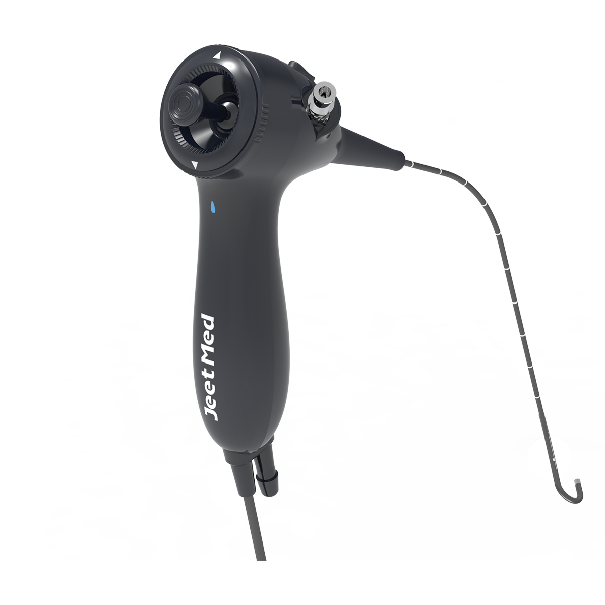
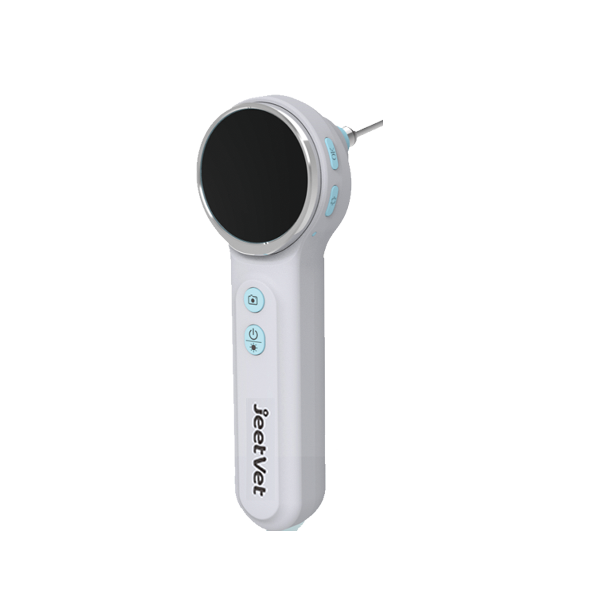
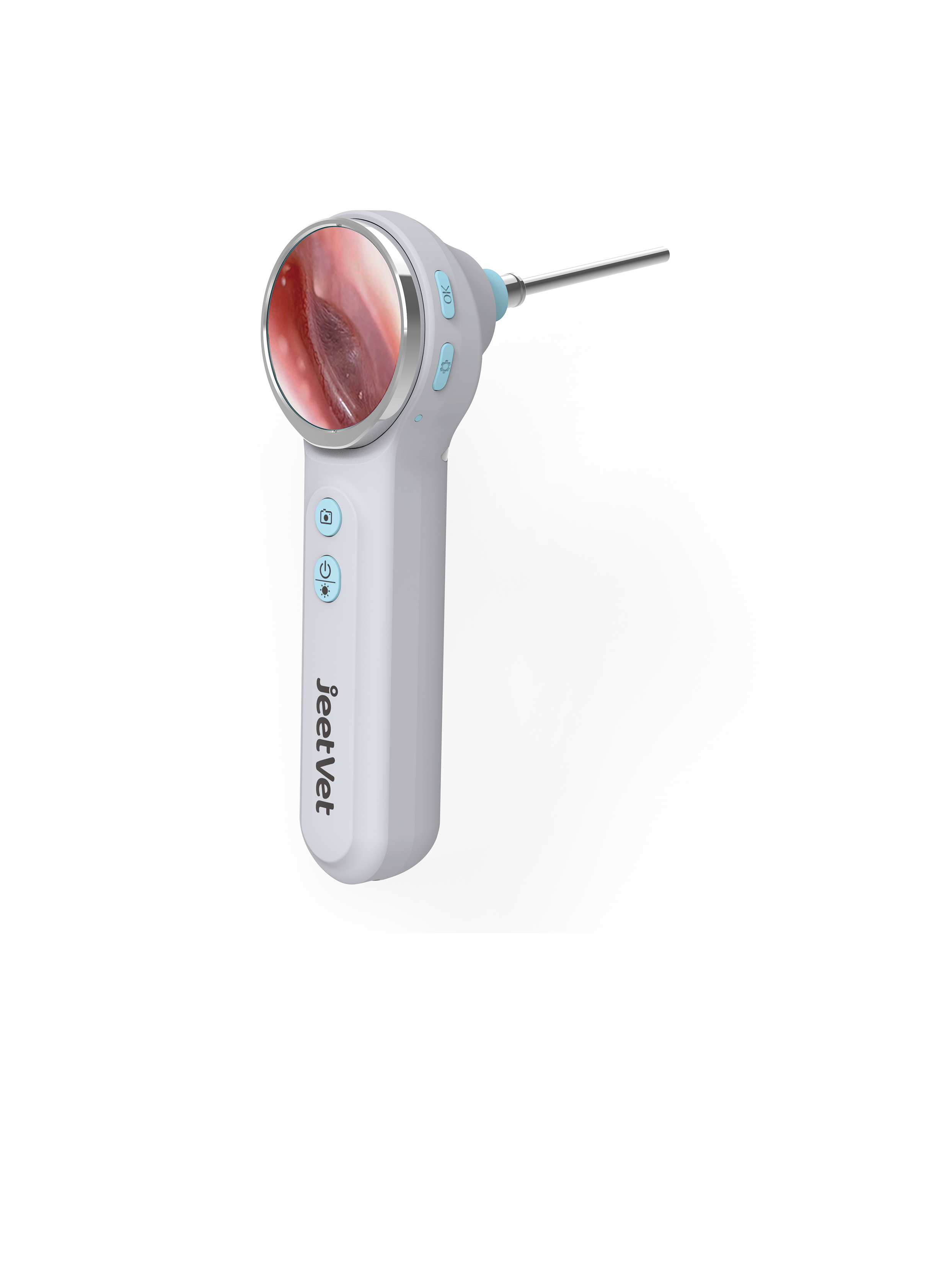
This guide works through two JeetVet instruments that cover different halves of that problem. The RAE-201 Veterinary Flexible Otoscope is built for animal ear and nasal examination, with a 600 mm working length, a 3.1 mm outer tube, a 1.2 mm inner channel, a 180° bending angle and a 120° field angle. The RAE-500 Portable Smart Veterinary Otoscope is a self-contained handheld with a 1.85-inch screen, a 1-megapixel camera, six LED lights, 1x, 1.2x and 1.5x image magnification, and 8 GB of built-in memory.
Problem Definition: Why Routine Otoscopy Stops Short
The dog and cat ear canal is not a straight tube. It runs vertically from the pinna and then turns horizontally toward the tympanic membrane, and a large share of chronic disease sits in that horizontal portion — debris, exudate, masses, and foreign material such as grass awns. A handheld cone illuminates the vertical canal well and the horizontal canal poorly. The gap between what the practitioner can see and what is actually present is where most ear-related treatment failures begin.
Five specific limits show up repeatedly:
- Depth. A rigid cone examines the accessible canal. It cannot be negotiated around the bend to inspect the horizontal canal and the region of the tympanum under direct vision.
- Field of view versus working distance. Wide viewing angles are useful for orientation; narrow ones are useful for detail. A single fixed optic forces a compromise that varies by case.
- No documentation. Findings are transcribed from memory into the record after the fact, which weakens follow-up comparison on the next visit.
- The nasal cavity is out of reach. Nasal discharge or obstruction generally requires a flexible instrument to visualise the passage, and sample collection to identify the cause.
- Operator dependence. Any instrument that must be interpreted by one person in the room cannot be confirmed by a second pair of eyes, and cannot easily be explained to an owner.
The practical question for a clinic is therefore not whether otoscopy is useful. It is which instrument configuration removes the specific limitation that keeps appearing in that clinic's case log. That distinction — between a specification sheet and a purchase decision — is what the rest of this article addresses.
Industry Background: A Small Category That Is Formalising Quickly
Veterinary endoscopy is a narrow but steadily formalising equipment category. Straits Research values the global veterinary endoscopy market at approximately USD 290–300 million in 2024/2025, with projections above USD 586 million by 2034. Market Research Future places North America at over 34.48% of global revenue as of 2024. Those figures are not uniform across research houses — Mordor Intelligence and Data Insights Reports publish lower base-year estimates — so any single market number should be read as a directional reference rather than a budgeting figure.
Two structural trends shape how a practice evaluates an otoscope in 2026. The first is image capture. Technavio and Strategic Market Research identify AI-assisted diagnostics in endoscopy as a trend advancing at a CAGR of 25.7%, which moves storage and image quality into the specification conversation rather than leaving them as accessories. The second is patient population. NAPHIA reports over 5 million insured pets in North America as of 2024, and insured populations typically accept advanced diagnostic procedures earlier than uninsured ones.
On the supply side, the recognised international names in the category include Karl Storz, Olympus Corporation, Eickemeyer and Fujifilm Holdings, according to Mordor Intelligence. Portable-device specialists such as JeetVet compete on a different axis: single-purpose, screen-integrated handheld instruments rather than full tower systems. Optical expectations across the category are framed by the ISO 8600 series, in which ISO 8600-1 covers general requirements for medical endoscopes and ISO 8600-3 covers field of view and direction of view. For ear and nasal work specifically, the practical consequence is that declared optical figures — field angle, field of view, depth of field — are the parameters worth reading before the price.
Detailed Solution: Two Instruments, Two Halves of the Same Exam
RAE-201: the deep-canal and nasal instrument
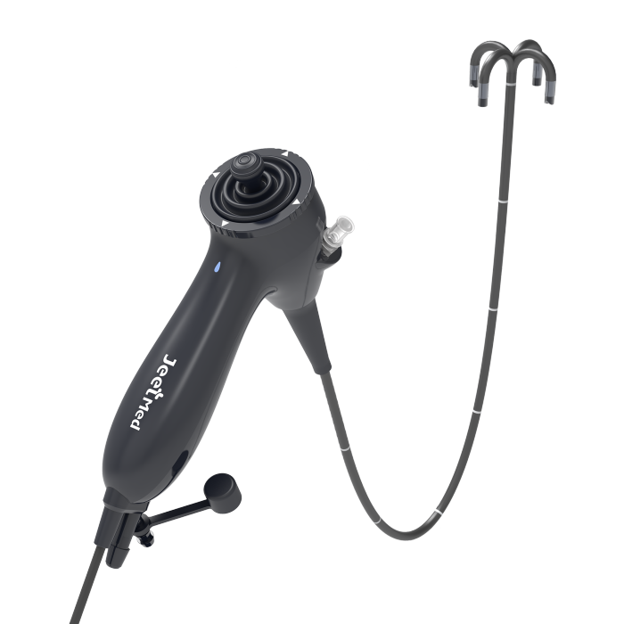
The RAE-201 is specified for animal ear and nasal examination, and its numbers are chosen for depth. A 600 mm working length is what makes a full-length examination of the canine ear canal and the nasal passage physically possible; the outer tube is only 3.1 mm across, which is what allows that length to enter a narrow canal without the insertion tube itself becoming the obstacle. The 1.2 mm inner channel is the instrument passage of the working assembly.
Two optical figures matter at the point of use. The 120° field angle gives wide orientation inside a curved canal — useful when the goal is to find the lumen rather than to inspect it. The 5–50 mm depth of field means the image stays in focus across the working range the practitioner actually uses, instead of requiring constant fine adjustment as the tip advances. Steering is up and down with a 180° bending angle, which is the motion needed to track the vertical-to-horizontal turn of the ear canal under direct vision rather than pushing blindly. Imaging output is rated at 160,000 pixels.
The ear-and-nose application profile published by the manufacturer lists the functions this configuration supports: aiding the removal of foreign bodies or masses, flushing the ear canal, and obtaining biopsies and cultures to identify the cause of nasal discharge or obstruction. In other words, the RAE-201 is the instrument in this pair that is expected to do interventional, sample-level work, not just visualisation.
RAE-500: the chairside, shareable instrument
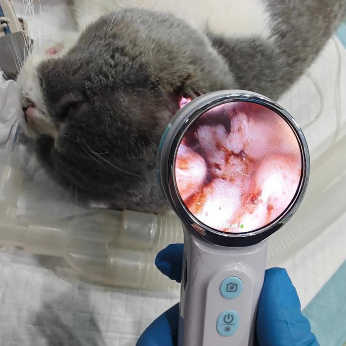
The RAE-500 answers a different constraint: speed and immediacy. Its defining feature is the 1.85-inch integrated screen. There is no external monitor to position, no cable to the practice laptop, and no setup step between the decision to examine and the examination itself. A 1-megapixel camera sits behind a 3.9 mm lens with a 78° field of view, six LED lights provide illumination inside the canal, and image or video resolution is 720 × 720 pixels.
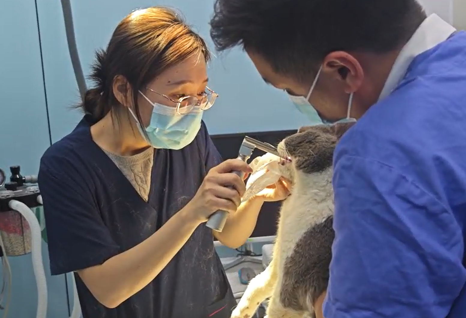
Magnification is selectable at 1x, 1.2x and 1.5x, and this is the feature that changes routine otoscopy most in daily use. At 1x the practitioner orients inside the canal. At 1.5x the magnification supports identification of small structures — mites, debris, small lesions, fine detail on the tympanic membrane. Because the image is on a screen rather than at an eyepiece, a technician, a colleague or an owner can look at the same finding at the same time and agree on what is being described.
Storage is built in at 8 GB, and the port is Type-C. Focal length is specified at 5–50 mm, the same near-range range the RAE-201 uses, which is appropriate for both instruments because both are working inside a canal rather than across a room. For practices moving toward image-based record keeping — the direction the AI-diagnostics trend points — the ability to capture and retain a 720 × 720 image of the canal without additional hardware is the practical benefit.
Shared environmental and handling specifications
Both instruments share the same declared operating envelope, which matters more in veterinary work than in human clinical settings because examinations happen outside purpose-built rooms. Operating temperature is 0°C to 40°C; storage temperature is −10°C to 55°C; relative humidity is ≤ 80% non-condensing. Continuous working mode is specified for clinical use, and the instruments are reusable after sterilisation. The manufacturer describes the intended environment as veterinary clinics, animal hospitals and field environments, supporting indoor and outdoor use, including mobile veterinary services. Material construction is stated as plastic, stainless steel, medical grade polymer and optical glass.
Those figures define where the equipment is expected to fail if handled carelessly. A unit left in a vehicle overnight in a cold climate, or a bag transported in high humidity without a desiccant, is outside the declared storage envelope and should be allowed to return to the operating range before use.
Step-by-Step Breakdown: Running an Ear and Nasal Examination
Step 1 — Choose the instrument by depth of the question, not by habit
Before restraint, decide what the examination has to answer. If the question is 'what is visible in the accessible canal', the RAE-500 is the faster route to an answer. If the question involves the full canal length, a suspected deep foreign body, a mass, or the nasal cavity, the RAE-201 is the correct starting instrument, because a surface examination that then requires a second procedure costs more time than starting deep.
Step 2 — Prepare and restrain with the canal axis in mind
The ear canal is straightened by pulling the pinna up and outward, and the instrument must follow that straightened axis. Restraint should hold the head stable in the line of the canal rather than merely holding the patient still. For deep work, the level of chemical restraint is a clinical decision, but it should be set before the insertion tube is advanced, not after the patient objects.
Step 3 — Start wide, then narrow
Begin at 1x on the RAE-500, or with the 120° field angle of the RAE-201, to locate the lumen. Orientation first, detail second. Advancing on a narrow view without first establishing where the canal runs is the most common way an insertion tube is pushed into a wall rather than along the passage.
Step 4 — Follow the canal with steering, not with force
The RAE-201 steers up and down with a 180° bending angle. The vertical-to-horizontal turn of the canine canal is negotiated by flexing the tip under direct vision, which is why the image should be watched continuously during advancement. Resistance at the tip is a signal to stop and re-orient, never a signal to push. The 5–50 mm depth of field means the image should remain readable across the working range; persistent blur usually indicates the tip is too close to tissue, not that the instrument needs adjustment.
Step 5 — Use magnification deliberately on the RAE-500
Switch to 1.2x or 1.5x once the region of interest is centred, not while searching. Magnification narrows the usable view, so it belongs after orientation. Six LED lights provide the illumination for this step, and the 720 × 720 capture records the finding at the moment of decision rather than in a note written afterwards.
Step 6 — Move to the nasal passage with the flexible instrument
Nasal examination follows the same principle as deep ear work: confirm the lumen before advancing, and keep the image under observation throughout. Where discharge or obstruction is present, the manufacturer's ear-and-nose application profile lists biopsy and culture collection as supported functions to identify the cause, and ear canal flushing as a further supported procedure. Capture or record the finding, then follow the manufacturer's cleaning and sterilisation instructions before the instrument is returned to storage.
Use Cases: Where Each Configuration Fits
Small animal clinics with a high otitis caseload
A clinic seeing repeated ear cases benefits most from documentation. The RAE-500 captures a 720 × 720 image into 8 GB of built-in memory and shows it on the integrated screen, so the finding at the first visit can be compared with the finding at the recheck. Magnification at 1.5x supports identification of fine detail that a routine cone view does not resolve.
Cases that need depth or sampling
Where the canal must be inspected to full length, where a foreign body is suspected deep in the horizontal canal, or where nasal discharge requires sampling to identify the cause, the RAE-201's 600 mm working length and 3.1 mm tube are the relevant capability. Its 1.2 mm inner channel is part of the working assembly that supports the flushing and sample-collection functions listed in the ear-and-nose application profile.
Mobile and field veterinary services
Both instruments are specified for use in veterinary clinics, animal hospitals and field environments, with indoor and outdoor use supported and an operating range of 0°C to 40°C. For mobile practice, the RAE-500 removes the need to unpack and position a display before examining, and the RAE-201 removes the need to transport a patient to a fixed tower for deep work. The storage range of −10°C to 55°C sets the limit for transport between visits.
Exotic and avian work
The category's application profile also covers avian and exotic animal endoscopy, where instrument diameter is the binding constraint. A 3.1 mm outer tube is the relevant figure in that context, because patient size, not examiner preference, determines whether an instrument can be used at all.
RAE-201 vs RAE-500: Published Specification Comparison
The table below uses only specifications published by the manufacturer. Where a parameter is not published for a model, it is marked as such rather than inferred.
| Specification | RAE-201 Veterinary Flexible Otoscope | RAE-500 Portable Smart Veterinary Otoscope |
|---|---|---|
| Purpose (published) | Animal ear and nasal examination | Portable smart veterinary otoscopy |
| Working length | 600 mm | Not published |
| Tube / lens diameter | 3.1 mm outer diameter of the tube | 3.9 mm diameter of lens |
| Working channel | 1.2 mm inner diameter of the channel | Not published |
| Steering / bending | 180° bending angle, up and down steering | Handheld; not applicable |
| Field angle / field of view | 120° field angle | 78° field of view |
| Depth of field / focal length | 5 mm – 50 mm depth of field | 5–50 mm focal length |
| Imaging | 160,000 pixels | 1 MP camera, 720 × 720 image / video |
| Display | Not published | 1.85-inch integrated screen |
| Magnification | Not published | 1x, 1.2x, 1.5x |
| Illumination | Not published | 6 LED lights |
| Storage and ports | Not published | Built-in 8 GB memory; Type-C port |
| Working environment | Veterinary clinics, animal hospitals, field environments, mobile veterinary services; indoor and outdoor use | Same declared environment and indoor / outdoor use |
| Operating / storage temperature | 0°C – 40°C operating; −10°C – 55°C storage | 0°C – 40°C operating; −10°C – 55°C storage |
| Best suited to | Deep canal inspection and nasal examination, flushing and sample collection | Fast chairside examination, magnification, and shared viewing or recording |
FAQ: Compliance, Capability, Budget, Evaluation and Delivery
1. Do these veterinary otoscopes have to meet a recognised optical standard?
For medical endoscopes, the ISO 8600 series is the widely referenced framework: ISO 8600-1 covers general requirements and ISO 8600-3 covers field of view and direction of view. Both JeetVet instruments declare their optical figures openly — the RAE-201 at a 120° field angle and the RAE-500 at a 78° field of view — which is what allows a buyer to check them against the standard's reporting expectations. On the manufacturing side, Jeet Medical operates under an ISO 13485 quality management system certified by SGS, supported by a 100,000-class cleanroom and a 10,000-class microbiological laboratory. Because certificate scope is defined per site and per product family, buyers should confirm the exact scope that applies to their market and their intended use before purchase.
2. Can a single otoscope handle both deep ear canal work and quick screening?
No single configuration is optimal for both. The RAE-201 is the deep instrument: 600 mm working length, 3.1 mm outer tube, 1.2 mm inner channel, 180° up-and-down steering, 120° field angle and a 5–50 mm depth of field. The RAE-500 is the fast instrument: a 3.9 mm lens, 78° field of view, 5–50 mm focal length, 1x / 1.2x / 1.5x magnification and a 1.85-inch screen with 8 GB of memory. Practices that see both routine otitis and deeper nasal or canal cases typically need the two capabilities rather than one compromise instrument.
3. What drives the cost difference between the two instruments anyway?
The cost drivers are structural rather than commercial. A flexible instrument with a steering mechanism, a bending section and a 1.2 mm working channel contains more precision mechanical assembly than a fixed handheld unit. Conversely, the RAE-500 carries the cost of an integrated 1.85-inch display, a 1 MP camera, six LED lights and 8 GB of built-in storage — but it removes the external monitor, capture hardware and cabling that a screenless optical system would otherwise require. For a small clinic, the relevant comparison is total installed setup against the case types actually seen, not a unit price against a unit price.
4. How should a practice evaluate either instrument before committing?
Evaluation should run against the clinic's own case mix. Count the ear and nasal cases from the last quarter, then map them onto the two configurations: how many needed deep canal access, how many needed nasal sampling, how many needed a second person to see the finding at the same time, and how many needed magnification for fine detail. Request the product documentation, including the published specification sheet and brochure, and discuss the evaluation approach with the manufacturer so the trial reflects real cases. JeetVet product information and the current catalogue can be reviewed at www.jeetvet.com, or requested directly through the contact channels listed at the end of this article.
5. What affects delivery timing once an order is placed?
Lead time is not a fixed published parameter for these models, because it depends on configuration, order quantity, sterilisation-related packaging and the documentation required for the destination market. The practical approach is to confirm the specific configuration and destination requirements with the supplier before the order is placed, and to request the delivery schedule in writing as part of the quotation. Buyers serving multiple markets should raise documentation requirements at the enquiry stage rather than after confirmation.
Conclusion: Match the Instrument to the Question
The RAE-201 and the RAE-500 do not compete for the same examination. The 600 mm working length, 3.1 mm outer tube and 180° up-and-down steering of the RAE-201 are what make full-length ear canal inspection, nasal visualisation and sample collection physically possible in dogs and cats. The 1.85-inch screen, 1-megapixel camera, six LED lights and 1x / 1.2x / 1.5x magnification of the RAE-500 are what make routine otoscopy fast, magnified and shareable at the point of care, with a 720 × 720 image stored in 8 GB of memory instead of a description written from memory. Both are specified for clinics, animal hospitals and field or mobile environments, with indoor and outdoor use, an operating range of 0°C to 40°C and a storage range of −10°C to 55°C.
Both instruments are manufactured by Jeet Medical (Shenzhen) Co., Ltd., traded under the JeetVet brand, founded in 2020 and operating a 5,000 m² facility with 130 employees, an annual output of 10,000 units, a 30-person R&D team and an export ratio of 50% across Europe, North America, Southeast Asia, Latin America and the Middle East. Manufacturing is certified under ISO 13485 by SGS.
Next step: match the specification to your caseload
Review the full RAE-201 and RAE-500 specifications in the JeetVet product catalogue, or request a quotation and sample evaluation for your clinic's case mix. Distributor and clinic enquiries are both welcome.
Download the JeetVet product brochure (PDF)
Website: www.jeetvet.com · Blog: blog.jeetvet.com
Email: troy@jeetmed.com · jeetvet66@gmail.com
Tel: +86 755-23251842 · +86 13381945549
WhatsApp: 8613816158380 · WeChat: 13381945549
Address: 9th Floor, Block B, Building 1, No. 76 Baohe Avenue, Longgang District, Shenzhen, China
Have Questions or Need More Details?
Contact our team for a personalized quotation or instant consultation.
Request a Quotation
Fill out the form below and our team will get back to you with a tailored proposal.
WhatsApp Direct Chat
Prefer to chat in real-time? Message us on WhatsApp for instant assistance & quick answers.
- Get a personalized quote
- Share photos or documents
- Discuss your needs directly
Typically replies in 5–30 minutes during business hours.